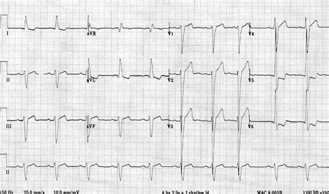
lv hypertrophy on ecg | left ventricular hypertrophy with repolarization abnormality lv hypertrophy on ecg The left ventricle hypertrophies in response to pressure overload secondary to conditions such as aortic stenosis and hypertension. This results in increased R wave . LV LT EE. Darba laiks Mājai Dārzam Sākam ar palodzi Remontam Būvei Atpūtai Pārtika Zoo Auto Velo Tehnika Pakalpojumi Ziedi Darbs DEPO DEPO online Darām kopā Mediju telpa. Darba laiks; Rīga, Kurzemes prospekts 3b; Rīga, Kurzemes prospekts 3b. Darba laiks Karte Mājai Dārzam . Darbs DEPO Darām kopā Sīkdatnes Privātuma politika .
0 · what is lvh on ecg
1 · signs of lvh on ecg
2 · lvh with repolarization abnormality ecg
3 · lv hypertrophy ecg criteria
4 · left ventricular hypertrophy with repolarization abnormality
5 · left ventricular hypertrophy life in the fast lane
6 · ecg showing lvh
7 · ecg in left ventricular hypertrophy
To delete a logical volume you need to first make sure the volume is unmounted, and then you can use lvremove to delete it. You can also remove a volume group once the logical volumes have been deleted and a physical volume after the volume group is deleted.
The left ventricle hypertrophies in response to pressure overload secondary to conditions such as aortic stenosis and hypertension. This results in increased R wave .The ECG Made Practical 7e, 2019; Kühn P, Lang C, Wiesbauer F. ECG Mastery: .ECG Pearl. There are no universally accepted criteria for diagnosing RVH in .Also known as: Left Atrial Enlargement (LAE), Left atrial hypertrophy (LAH), left .
what is lvh on ecg
signs of lvh on ecg
The delay between activation of the RV and LV produces the characteristic “M .The ECG Made Practical 7e, 2019; Kühn P, Lang C, Wiesbauer F. ECG Mastery: .Kühn P, Lang C, Wiesbauer F. ECG Mastery: The Simplest Way to Learn the .
The most common causes of left ventricular hypertrophy are aortic stenosis, aortic regurgitation, hypertension, cardiomyopathy and coarctation of the aorta. .
The left ventricle hypertrophies in response to pressure overload secondary to conditions such as aortic stenosis and hypertension. This results in increased R wave amplitude in the left-sided ECG leads (I, aVL and V4-6) and increased S .The most common causes of left ventricular hypertrophy are aortic stenosis, aortic regurgitation, hypertension, cardiomyopathy and coarctation of the aorta. There are several ECG indexes, which generally have high diagnostic specificity but low sensitivity. Left ventricular hypertrophy (LVH) refers to an increase in the size of myocardial fibers in the main cardiac pumping chamber. Such hypertrophy is usually the response to a chronic pressure or volume load. The two most common pressure overload states are systemic hypertension and aortic stenosis.
romaorologi orologi rolex usati via cavour roma
lvh with repolarization abnormality ecg

rubato rolex attore gandolfini
According to the American Society of Echocardiography and/European Association of Cardiovascular Imaging, LVH is defined as an increased left ventricular mass index (LVMI) to greater than 95 g/m in women and increased LVMI to greater than 115 g/m in men. Electrocardiogram. Also called an ECG or EKG, this quick and painless test measures the electrical activity of the heart. During an ECG, sensors called electrodes are attached to the chest and sometimes to the arms or legs. Wires connect the sensors to a machine, which displays or prints results. Uncontrolled high blood pressure is the most common cause of left ventricular hypertrophy. Complications include irregular heart rhythms, called arrhythmias, and heart failure. Treatment of left ventricular hypertrophy depends on the cause. Treatment may include medications or surgery. The ECG changes in a patient with left ventricular hypertrophy (LVH) were described 117 years ago by Einthoven in 1906 (Einthoven, 1957). He drew attention to the distinctive finding—the increased QRS amplitude in the “left hand to left foot lead” (i.e., lead III).
Left ventricular hypertrophy can be diagnosed on ECG with good specificity. When the myocardium is hypertrophied, there is a larger mass of myocardium for electrical activation to pass.
Left ventricular hypertrophy (LVH) refers to an increase in the size of myocardial fibers in the main cardiac pumping chamber. Such hypertrophy is usually the response to a chronic pressure or volume load. The two most common pressure overload states are .Current electrocardiographic (ECG) criteria for the diagnosis of left ventricular hypertrophy (LVH) have low sensitivity. Objectives: The goal of this study was to test a new method to improve the diagnostic performance of the electrocardiogram. The left ventricle hypertrophies in response to pressure overload secondary to conditions such as aortic stenosis and hypertension. This results in increased R wave amplitude in the left-sided ECG leads (I, aVL and V4-6) and increased S .
The most common causes of left ventricular hypertrophy are aortic stenosis, aortic regurgitation, hypertension, cardiomyopathy and coarctation of the aorta. There are several ECG indexes, which generally have high diagnostic specificity but low sensitivity. Left ventricular hypertrophy (LVH) refers to an increase in the size of myocardial fibers in the main cardiac pumping chamber. Such hypertrophy is usually the response to a chronic pressure or volume load. The two most common pressure overload states are systemic hypertension and aortic stenosis. According to the American Society of Echocardiography and/European Association of Cardiovascular Imaging, LVH is defined as an increased left ventricular mass index (LVMI) to greater than 95 g/m in women and increased LVMI to greater than 115 g/m in men.
lv hypertrophy ecg criteria
Electrocardiogram. Also called an ECG or EKG, this quick and painless test measures the electrical activity of the heart. During an ECG, sensors called electrodes are attached to the chest and sometimes to the arms or legs. Wires connect the sensors to a machine, which displays or prints results.
Uncontrolled high blood pressure is the most common cause of left ventricular hypertrophy. Complications include irregular heart rhythms, called arrhythmias, and heart failure. Treatment of left ventricular hypertrophy depends on the cause. Treatment may include medications or surgery.
The ECG changes in a patient with left ventricular hypertrophy (LVH) were described 117 years ago by Einthoven in 1906 (Einthoven, 1957). He drew attention to the distinctive finding—the increased QRS amplitude in the “left hand to left foot lead” (i.e., lead III).
Left ventricular hypertrophy can be diagnosed on ECG with good specificity. When the myocardium is hypertrophied, there is a larger mass of myocardium for electrical activation to pass.Left ventricular hypertrophy (LVH) refers to an increase in the size of myocardial fibers in the main cardiac pumping chamber. Such hypertrophy is usually the response to a chronic pressure or volume load. The two most common pressure overload states are .
scandalo dei rolex

Buy Dell 4GB DDR3 SDRAM Memory Module. 4GB 1600MHZ NON-ECC DDR3 SDRAM 240PIN UBDIMM F/OPTI 3010 7010 9010. 4 GB (1 x 4 GB) - DDR3 SDRAM - 1600 MHz DDR3-1600/PC3-12800 - Non-ECC - Unbuffered - 240-pin DIMM: Memory - Amazon.com FREE DELIVERY possible on eligible purchases
lv hypertrophy on ecg|left ventricular hypertrophy with repolarization abnormality


