lv clot on echo | Lv thrombus heart attack lv clot on echo CMR may be most appropriate (1) when there is the suggestion of a possible LV thrombus on echocardiogram but echocardiography imaging (even with an ultrasound . Fotogrāfs Edgars Pohevičs. Tavs dzīves mirkļu fotogrāfs. Previous Slide Next Slide. Kāzu foto. Kāzu Foto no gatavošanās līdz mičošanai, vairāk info par kāzām lasi manā kāzu fotogrāfa lapā SPIED ŠEIT. Pakalpojumā ietilpst: Kāzu fotografēšana līdz 14 stundām – no līgavas/līgavaiņa gatavošanās līdz mičošanai (no .
0 · treatment for Lv thrombus
1 · left ventricular thrombus risk assessment
2 · left ventricular thrombus formation symptoms
3 · left ventricular thrombus
4 · echocardiography for Lv thrombus
5 · Lv thrombus risk management
6 · Lv thrombus recurrence rate
7 · Lv thrombus heart attack
Fathom LV Specs and Features. Structure: Rigid / Hard Shell. Cockpit Type: Sit Inside. Seating Configuration: Solo. Ideal Paddler Size: Average Adult, Larger Adult. Skill Level: Intermediate, Advanced. Additional Attributes. Optional installed rudder. Eddyline Kayaks. Fathom LV Reviews. Submit Your Review.
Left ventricular (LV) thrombus may develop after acute myocardial infarction (MI) and occurs most often with a large, anterior ST-elevation MI (STEMI). However, the use of reperfusion therapies, including percutaneous coronary intervention and fibrinolysis, has .Authors Gregory YH Lip, MD, FRCPE, FESC, FACC Price-Evans Professor of .Echocardiographic Algorithm for Post-Myocardial Infarction LV Thrombus: A .
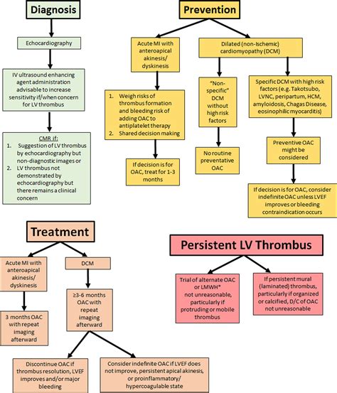
CMR may be most appropriate (1) when there is the suggestion of a possible LV thrombus on echocardiogram but echocardiography imaging (even with an ultrasound .
LV regional wall akinesia and dyskinesia result in blood stasis, often recognised on two dimensional echocardiography by the occurrence of spontaneous LV contrast. Prolonged . Left ventricular (LV) thrombus may develop after acute myocardial infarction (MI) and occurs most often with a large, anterior ST-elevation MI (STEMI). However, the use of reperfusion therapies, including percutaneous coronary intervention and fibrinolysis, has significantly reduced the risk. CMR may be most appropriate (1) when there is the suggestion of a possible LV thrombus on echocardiogram but echocardiography imaging (even with an ultrasound enhancing agent) is not diagnostic and (2) when echocardiography does not demonstrate LV thrombus but a clinical concern (eg, cardioembolic stroke) remains.
LV regional wall akinesia and dyskinesia result in blood stasis, often recognised on two dimensional echocardiography by the occurrence of spontaneous LV contrast. Prolonged ischaemia leads to subendocardial tissue injury with inflammatory changes.We identified patients with LV thrombus on echocardiography (with and without contrast) at Brigham and Women’s Hospital between January 2008 and May 2015. Etiologies, treatment strategies, follow-up imaging, and 1-year outcomes were recorded after physician chart review. Standard transthoracic echocardiography (TTE) is typically the screening modality of choice for LV thrombus detection and should be performed within 24 hours of admission in those at high risk for apical LV thrombus (e.g., those with large or anterior MI or those receiving delayed reperfusion).Accurate detection of left ventricular (LV) thrombus is important, as thrombus provides a substrate for thromboembolic events and a rationale for anticoagulation. Non-contrast echocardiography (echo) detects LV thrombus based on anatomical appearance.
Left ventricular thrombus is a blood clot in the left ventricle of the heart. LVT is a common complication of acute myocardial infarction (AMI). [1] [2] Typically the clot is a mural thrombus, meaning it is on the wall of the ventricle. [3]
treatment for Lv thrombus

Indications for LVAD. Bridge to transplant. Bridge to recovery: Acute myocarditis, Tako Tsubo, Post MI Shock. Destination Therapy: Refractory HF, not transplant candidate. Anatomy of an LVAD. Inflow Cannula (LV) Pump: Axial magnetic Rotor (HMII) Centrifugal propeller (HVAD) On the basis of limited data, patients with nonischemic cardiomyopathy with LV thrombus should be treated with OAC for at least 3–6 months, with discontinuation if LV ejection fraction improves to >35% (assuming resolution of the LV thrombus) or if major bleeding occurs.
rolex 40mm sd vs submarier
Among patients with possible LVT as the clinical indication for echo, sensitivity and positive predictive value were markedly higher (60%, 75%). Regarding sensitivity, echo performance related to LVT morphology and mirrored cine-CMR, with protuberant thrombus typically missed when small (p≤0.02). Left ventricular (LV) thrombus may develop after acute myocardial infarction (MI) and occurs most often with a large, anterior ST-elevation MI (STEMI). However, the use of reperfusion therapies, including percutaneous coronary intervention and fibrinolysis, has significantly reduced the risk. CMR may be most appropriate (1) when there is the suggestion of a possible LV thrombus on echocardiogram but echocardiography imaging (even with an ultrasound enhancing agent) is not diagnostic and (2) when echocardiography does not demonstrate LV thrombus but a clinical concern (eg, cardioembolic stroke) remains.
LV regional wall akinesia and dyskinesia result in blood stasis, often recognised on two dimensional echocardiography by the occurrence of spontaneous LV contrast. Prolonged ischaemia leads to subendocardial tissue injury with inflammatory changes.We identified patients with LV thrombus on echocardiography (with and without contrast) at Brigham and Women’s Hospital between January 2008 and May 2015. Etiologies, treatment strategies, follow-up imaging, and 1-year outcomes were recorded after physician chart review. Standard transthoracic echocardiography (TTE) is typically the screening modality of choice for LV thrombus detection and should be performed within 24 hours of admission in those at high risk for apical LV thrombus (e.g., those with large or anterior MI or those receiving delayed reperfusion).
left ventricular thrombus risk assessment
Accurate detection of left ventricular (LV) thrombus is important, as thrombus provides a substrate for thromboembolic events and a rationale for anticoagulation. Non-contrast echocardiography (echo) detects LV thrombus based on anatomical appearance.Left ventricular thrombus is a blood clot in the left ventricle of the heart. LVT is a common complication of acute myocardial infarction (AMI). [1] [2] Typically the clot is a mural thrombus, meaning it is on the wall of the ventricle. [3]
Indications for LVAD. Bridge to transplant. Bridge to recovery: Acute myocarditis, Tako Tsubo, Post MI Shock. Destination Therapy: Refractory HF, not transplant candidate. Anatomy of an LVAD. Inflow Cannula (LV) Pump: Axial magnetic Rotor (HMII) Centrifugal propeller (HVAD) On the basis of limited data, patients with nonischemic cardiomyopathy with LV thrombus should be treated with OAC for at least 3–6 months, with discontinuation if LV ejection fraction improves to >35% (assuming resolution of the LV thrombus) or if major bleeding occurs.

left ventricular thrombus formation symptoms


rolex 6239 cherry logo
rolex 4471
Fans attending EDC Las Vegas in 2024 can take in performances by Martin Garrix, Eric Prydz, Zedd, Alison Wonderland, Kaskade, John Summit, Dom Dolla, deadmau5, Alesso, Armin van Buuren, ILLENIUM,.
lv clot on echo|Lv thrombus heart attack


